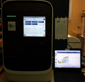
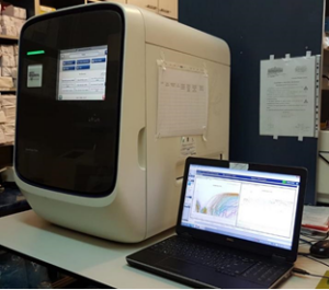
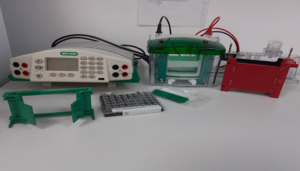
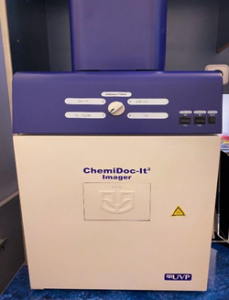
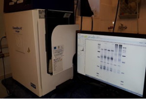
Our laboratory carries out its activity in two premises. Our preparation rooms are organised as to ensure the correct flow of reactions, avoiding contamination.

The cell biology facility is equipped with 4 biosafety cabinets (s@feflow 1.8 Bioair; Holten LaminairA), 6 incubators (Thermo Scientific; Memert; IGO 150 Jouan) and centrifuges. For cutting-edge microscopy and the support of diverse image acquisition and analysis methods, a Nikon Eclipse Ti-E is employed.

For activities such as cell culture, viability analysis and for demonstration and teaching programs we use a Nikon Eclipse TS100 inverted microscope.

Furthermore, the xCELLigence RTCA system provides relevant data on cell proliferation, adhesion, spreading, cell invasion, and migration by measuring the electrical impedance of the cell layer in real time.  For the capture of in-focus images, multi-sample, and multi-point imaging the BioStation IM system allows us to create a cell friendly environment and a simple long-term time-lapse data acquisition.

The molecular biology area of our laboratory is equipped with innovative cutting-edge tools prepared for the complex analysis of MRD and chimerism. In the past years, we were able to acquire and also to make the transition from classical polymerase chain reaction (PCR) (Mastercycler Nexus GX2-Eppendorf) using gel electrophoresis to Real-Time quantitative PCR (RT-qPCR; QuandStudio 7 Flex Real-Time System-Thermo Fisher Scientifichttps://www.thermofisher.com/ro/en/home/life-science/pcr/real-time-pcr/real-time-pcr-instruments/quantstudio-7-flex-real-time-pcr-system.html


and the recent droplet digital PCR (dd-PCR) technique (QX200 Digital Droplet PCR SystemBioRadhttps://www.bio-rad.com/en-us/product/qx200-droplet-digital-pcr-system?ID=MPOQQE4VY

Having both methods (RT-qPCR versus dd-PCR) at hand, we were able to perform numerous comparative studies in terms of specificity and sensitivity which is a steppingstone in molecular diagnostics. After hematopoietic transplant, the mixed chimerism assessment is critical for detecting early signs of graft rejection, preventing relapse of the underlying disease, and monitoring of MRD. We explore the advantages of using INDEL detection by PCR to increase the sensitivity of the method. The equipment was acquired through POS-CCE 2.2.1. Cod SMIS CSNR 1882/49159 (CAMED).

For identification of specific proteins from a complex mixture extracted from tissues or cell culture, our laboratory employs the western blot method (Biorad system). Protein detection and visualization on the membrane is acquired with UVP’s ChemiDoc-It2.

ro_RORomanian